A female octopus (Haliphron atlanticus) consumes a jellyfish at 800 meters depth. Observing feeding interactions helps scientists understand how midwater communities function and offers insights into processes we all depend on, such as carbon cycling through the largest habitat on Earth. Credit: ROV SuBastian/Schmidt Ocean Institute
An expedition to international waters off the coast of Brazil used state-of-the-art imaging systems to confirm new midwater species and observed the living 3D cellular structure of a microbe—a first for seagoing research
An international team of midwater experts on board Schmidt Ocean Institute’s research vessel Falkor (too) discovered over two dozen new marine species on a recent expedition off the coast of Brazil in the tropical South Atlantic Ocean. The scientists used advanced technologies to explore the ocean’s midwater—the water between the sunlit layer and the seafloor—which is Earth’s largest and least explored habitable ecosystem. It can take scientists decades to identify and describe new species, but the combination of technology and expertise enabled the team to confirm these species as new within a matter of days.
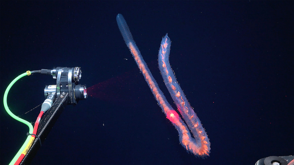
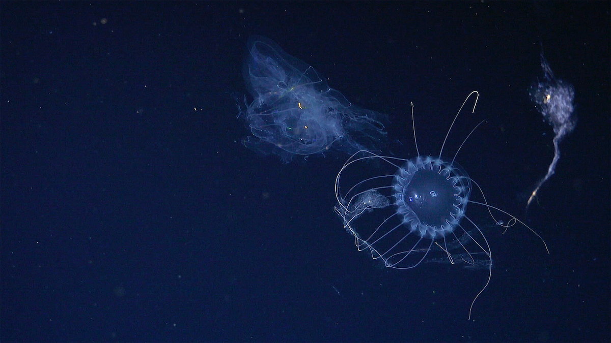
The list consists of an amphipod, a gossamer worm, nine jellyfish, seven siphonophores (colonial marine invertebrates related to jellyfish), seven ctenophores (comb jellies), four larvaceans (tadpole-like creatures that live in mucus houses and are more closely related to humans than invertebrates), and two giant rhizarians (single-celled organisms visible to the naked eye).
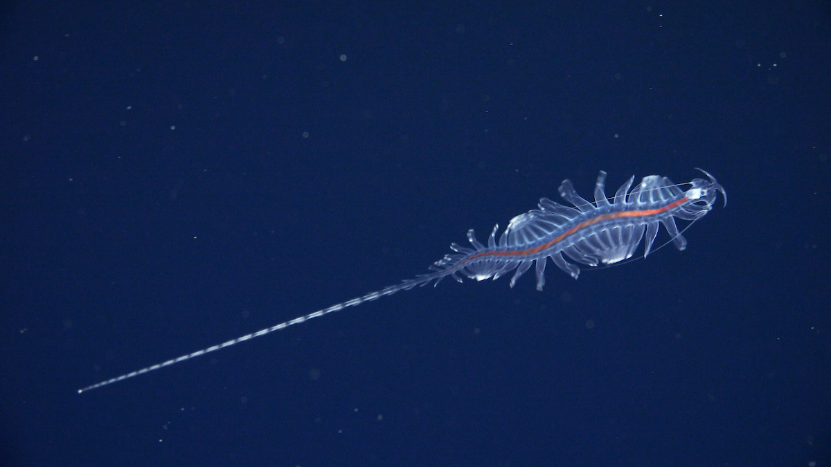
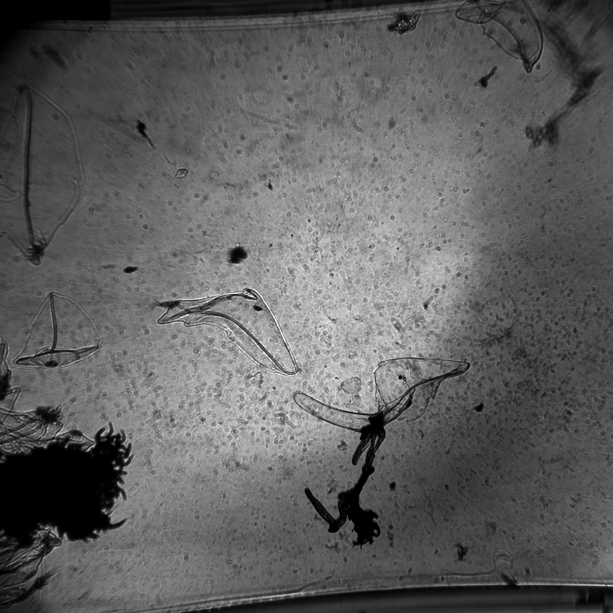
This is a new species from the genus Tomopteris, commonly known as gossamer worms. The expedition science team tested new technology that provides scientists with new, non-invasive ways to study these remarkable animals. Credit: ROV SuBastian/Schmidt Ocean Institute
“The largest habitat on Earth, the midwater, is filled with incredible animals we are only just starting to understand,” said the expedition’s chief scientist, Dr. Karen Osborn of the Smithsonian National Museum of Natural History. “I continue to be fascinated by the fantastic variety of solutions they have evolved to survive in this formidable environment, and that drives me to keep asking questions about our ocean.”
The team witnessed far more diversity and abundance of midwater organisms than they expected, said Osborn, including glass squid and a pelagic octopus feeding on a bright red jellyfish.
The ocean’s midwater is one of the most challenging areas on Earth to explore because of its inaccessibility and immense volume. The Sasakawa Peace Foundation’s Ocean Shot Research Grant Program funded two midwater programs that made this work possible, one based at the University of Western Australia and the other at Bigelow Laboratory for Ocean Sciences, USA.
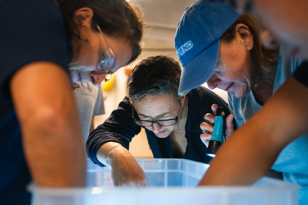
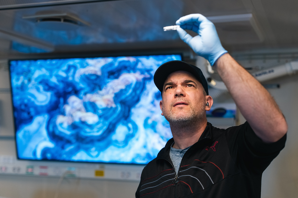
Expedition Chief Scientist Dr. Karen Osborn (Smithsonian National Museum of Natural History) works with Heather Judkins (University of South Florida, St. Petersburg) and Dr. Silvina Botta (Universidade Federal do Rio Grande) in the wet lab of R/V Falkor (too), gathering tiny translucent animals from a large container for further study. The team used cutting-edge technology both deep underwater and aboard the ship, including specialized microscopes, DNA sampling, stable isotope analysis, and multiple imaging systems. Credit: Alex Ingle/Schmidt Ocean InstitutePrincipal Investigator Dr. John Burns (Bigelow Laboratory for Ocean Sciences) examines a sample, which will be used for his genetic sequencing work in the main lab of R/V Falkor (too). In tandem with high-resolution imagery gathered at depth, the team sequenced genomes from collected specimens on board the vessel, enabling them to rapidly identify new species. Credit: Alex Ingle/Schmidt Ocean Institute
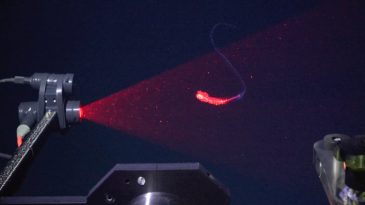
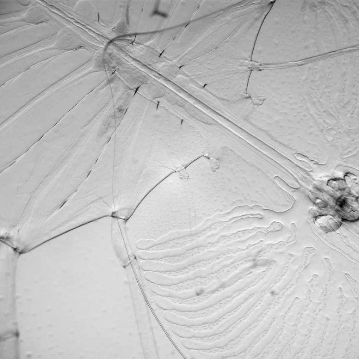
The technologies used to identify new species were a combination of imaging systems and genetic analyses. The imaging systems included the DeepPIV (particle image velocimetry) and EyeRIS (remote imaging system) instruments, developed by the Bioinspiration Lab at MBARI (Monterey Bay Aquarium Research Institute), which were attached to Schmidt Ocean Institute’s remotely operated vehicle (ROV) SuBastian. DeepPIV and EyeRIS are sophisticated, non-invasive tools for scanning marine animals; they use lasers to scan organisms and create 3D images of them. In addition, the team attached a shadowgraph camera from the Japan Agency for Marine-Earth Science and Technology (JAMSTEC) to the ROV, which can image the finer details of animals not visible in the 3D scans. The images help scientists describe the shape and internal structures of animals without having to collect them.
“It’s an incredible honor to not only view and experience this rare and inspiring midwater life, but also to be able to work towards describing and sharing that life broadly through the use of novel, non-invasive technologies,” said Dr. Kakani Katija, principal engineer of the Bioinspiration Lab at MBARI.
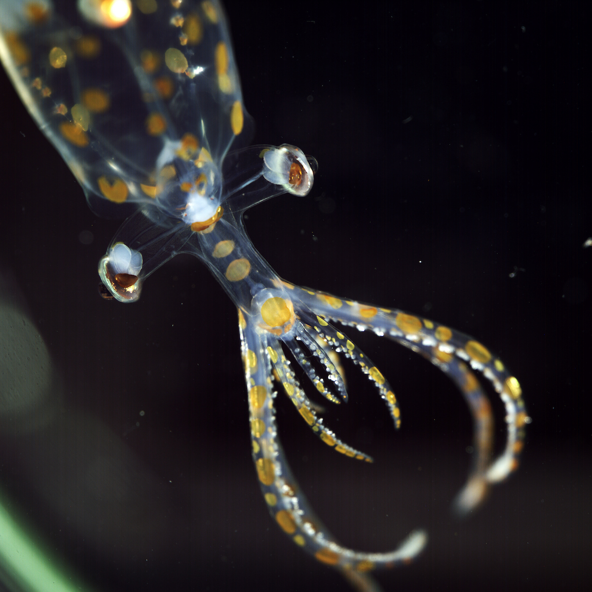
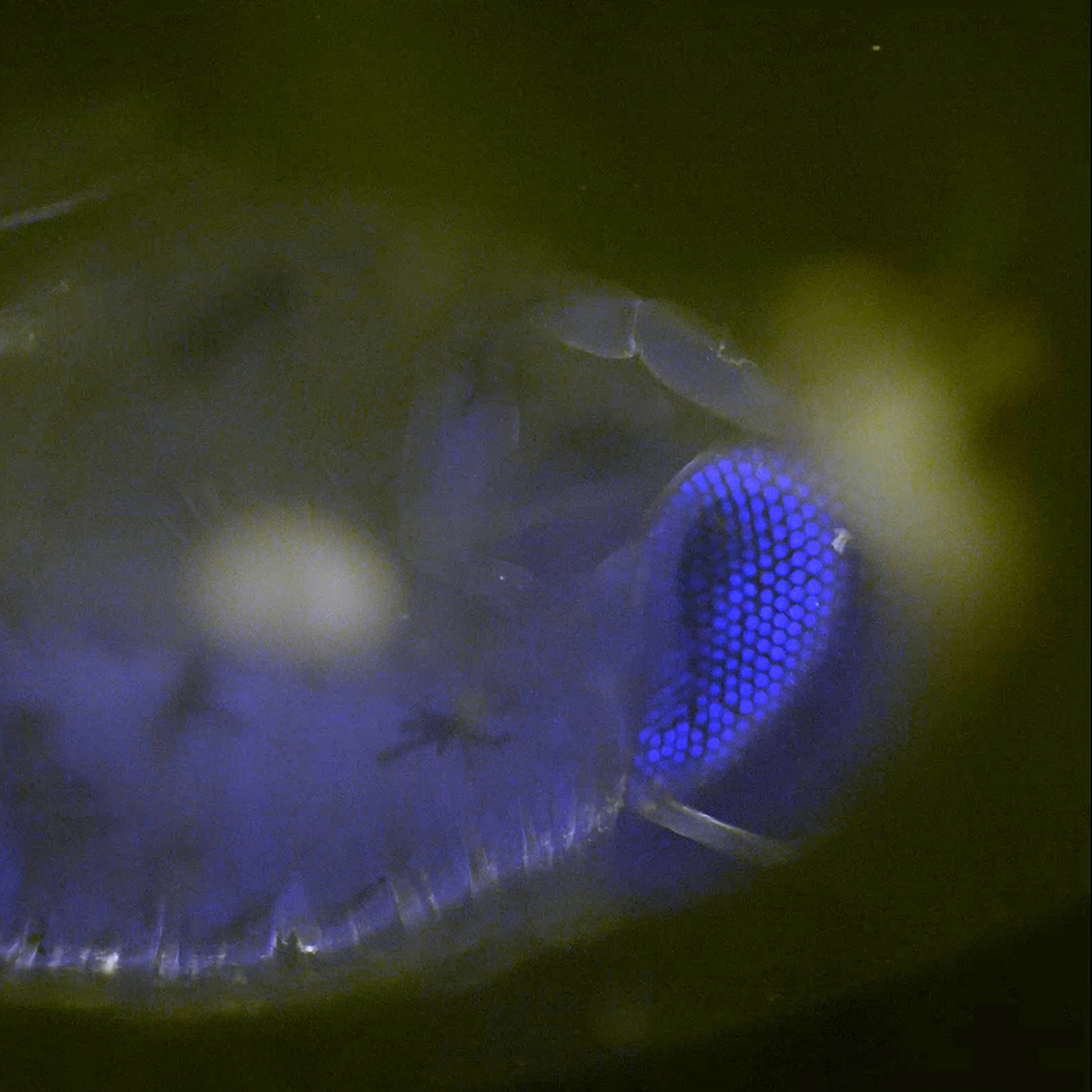
This juvenile glass squid, collected by ROV SuBastian at 779 meters depth in the South Atlantic, was photographed on R/V Falkor (too) using a prototype multiview macro camera system developed through a collaboration between the Dr. Jan Hemmi (University of Western Australia, the Bioinspiration Lab at MBARI and Dr. Karen Osborn (Smithsonian National Museum of Natural History). The system allows scientists on the ship to quickly document the finest details of an animal from three directions at once. This data gathering reduces the disturbance to the animal and captures anatomical, color and posture details that are lost within minutes to hours once the animal is collected. Credit: Emily Clark/MBARI via Schmidt Ocean InstituteThis image captured by a shadowgraph camera shows the front, side and top views of protective shield tissues from a siphonophore. Credit: Dhugal Lindsay/JAMSTECA microscopic view shows the complex, honeycomb-like structure of a hyperiid’s eye, a tiny shrimp-like crustacean that lives in the deep ocean. Researchers used advanced 3D confocal live-imaging technology to capture individual cone cells that form the creature’s compound eyes. Credit: Manu Prakash/Stanford UniversityA siphonophore is scanned using Deep Particle Image Velocimetry (DeepPIV) at a depth of 350 meters. This species was undescribed prior to this encounter and is likely new to science. DeepPIV is a laser- and optics-based imaging system that quantifies both the motion of liquids and the 3D shape of transparent animals. The imaging system was developed by the Bioinspiration Lab at MBARI (Monterey Bay Aquarium Research Institute) to create 3D models of gelatinous animals. In this image, DeepPIV is attached to the remotely operated vehicle (ROV) SuBastian. Credit: ROV SuBastian/Schmidt Ocean InstituteThis is a phase-contrast image of a live phyllosoma, a larval stage of a spiny or slipper lobster (magnified 10x). The microscopic details of the animal’s completely transparent digestive and nervous systems—tightly sandwiched between its ultra-thin, transparent exoskeleton—are visible in this image. Credit: Manu Prakash/Stanford UniversityThis jelly was collected at 1,157 meters depth by ROV SuBastian and photographed on board Falkor (too) using a prototype Multiview Macro Camera system. Credit: Emily Clark/MBARI
Many midwater animals are gelatinous, with soft, delicate bodies that are often damaged by traditional sampling methods. To address this challenge, the expedition used additional technologies that allowed scientists to observe animals in a controlled environment that mimics their natural habitat. These included a virtual reality chamber developed at the University of Western Australia and a “gravity machine” developed at Stanford University—a specialized microscope that functions as a hydrodynamic treadmill for studying microbes.
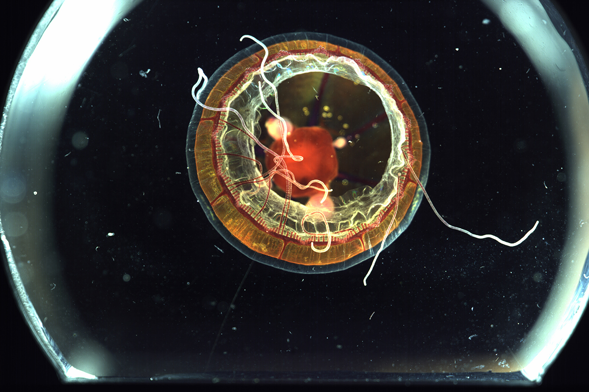
The team used another microscope developed at Stanford University to gain critical new insights into the physiology of midwater animals. The microscope, known as Squid, is an open-source, confocal microscope. Using Squid, the team achieved a first for research at sea and imaged living internal cellular structures in 3D. One of the organisms imaged was a large single-celled microbe called a protist. The microscope enabled the scientists to observe how the protist’s cellular structure interacted with its glass skeleton.
“This opens a new door for researching deep-sea physiology, linking cellular architectures to organism function. We can now witness live internal processes within these extreme organisms adapted to withstand immense pressure and darkness,” said Dr. Manu Prakash of Stanford University.
In tandem with the high-resolution imagery, the team sequenced genomes from collected specimens onboard the vessel, enabling them to rapidly identify new species under the leadership of Dr. Cheryl Ames of Tohoku University and Dr. John Burns of Bigelow Laboratory.
“The novel suite of technologies on this cruise is a glimpse into the future of marine biological science,” said Schmidt Ocean Institute’s Executive Director, Dr. Jyotika Virmani. “Schmidt Ocean Institute’s mission is to push technological advancement and this was our third cruise in collaboration with this team of scientists and engineers to test and further develop this innovative midwater equipment. We look forward to a future in which scientists study marine life as elegantly as this team did—and in virtual reality.”
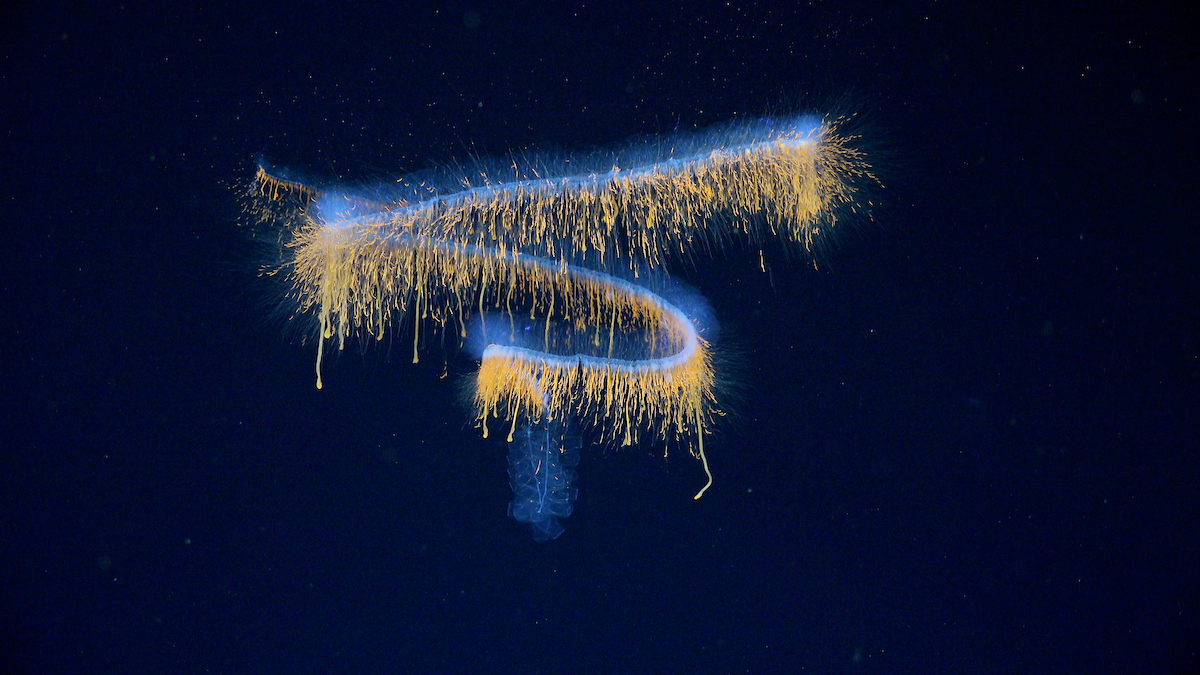
The team collected footage of this siphonophore at 552 meters depth. The imaging systems tested on R/V Falkor (too) allowed researchers to create millimeter-scale, 3D renderings of the creature in its natural habitat. Most species identifications take place ashore, using samples or small pieces, but these systems allow scientists to see and study the entire animal as it lives in the water. Based on images and measurements collected at sea, Dr. Dhugal Lindsay of JAMSTEC (Japan Agency for Marine-Earth Science and Technology) is confident that this animal belongs to an undescribed genus, perhaps even a new family of physonect siphonophores. Based on the detailed anatomical and genetic data collected in the water and on board, scientists will be able to compare this animal to those collected elsewhere around the globe and give this physonect a name. Credit: ROV SuBastian/Schmidt Ocean InstituteA siphonophore—a colonial marine invertebrate related to jellyfish—is scanned using Deep Particle Image Velocimetry (DeepPIV) at a depth of 930 meters. DeepPIV is attached to the remotely operated vehicle (ROV) SuBastian; it is a laser- and optics-based imaging system that quantifies both the motion of liquids and the 3D shape of transparent animals. The imaging system was developed by the Bioinspiration Lab at MBARI (Monterey Bay Aquarium Research Institute) to create 3D models of gelatinous animals. Credit: ROV SuBastian/Schmidt Ocean InstituteA Solmissus, or dinner plate jellyfish, preys upon a ctenophore, commonly known as a comb jelly. Unlike most jellyfish that passively drag their tentacles behind them, Solmissus swims with their tentacles extended in front of their body to snare ctenophores before vibrations alert the prey. They are believed to be gelatinous apex predators that play a major role in regulating comb jelly populations in the Ocean’s twilight and midnight zones. Credit: ROV SuBastian/Schmidt Ocean Institute


